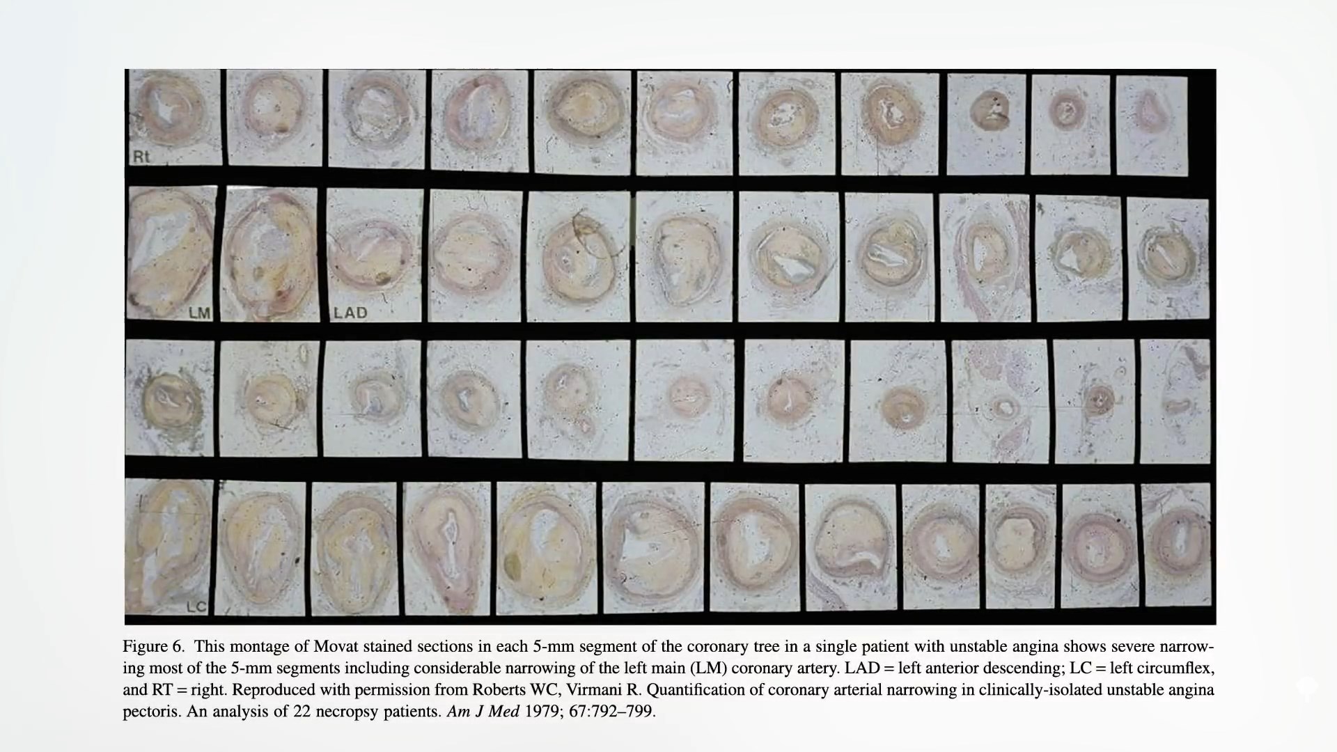
Most heart attacks are caused by non-obstructive plaques that penetrate the entire coronary tree. There is no such thing as “1-vascular disease”, “bivascular disease”, or “left main disease”. Atherosclerotic plaques are continuous throughout the coronary arteries of heart attack victims.
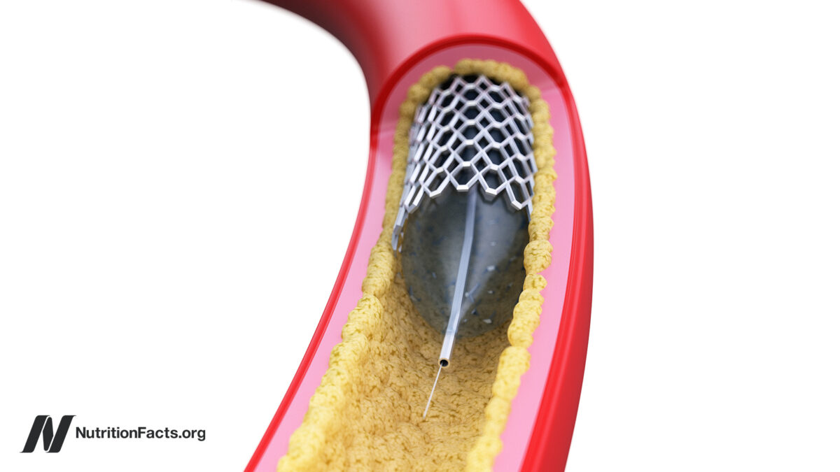
In angioplasty, small balloons are inserted into narrow coronary arteries, fed into the heart, and open more wider to improve blood flow. It was not tested in randomized controlled trials until 1992. Not only did they fail to prevent a heart attack, they were unable to show the benefits of survival. However, the researchers only followed patients for six months, including those with relatively mild illnesses that may not have been sufficiently ill to benefit from this procedure. Please enter your masking. The researchers registered people with a high severe obstruction in the left anterior descending coronary artery. Written or widow manufacturers (as coronary artery disease is also a number one killer for women) and lasted for years. Survey results? There was no difference in subsequent mortality or heart attack rates. However, the trial only had around 200 patients. Perhaps the benefits were so subtle that more patients were needed to tease the effects. Participate in a RITA-2 study that randomized more than 1,000 patients. Researchers certainly found a clear difference between future death and risk of heart attacks, but that was in the wrong direction. The angioplasty group took twice as much risk as the group randomized to abandon the surgery, 1:18 for the reason angioplasty stents do not function well, as shown below.
But this was before stents became popular. How about inserting a stent (metal mesh tube) permanently to open the artery, as you can see in my video, as well as stents (metal mesh tubes)? Certainly, it has to help.
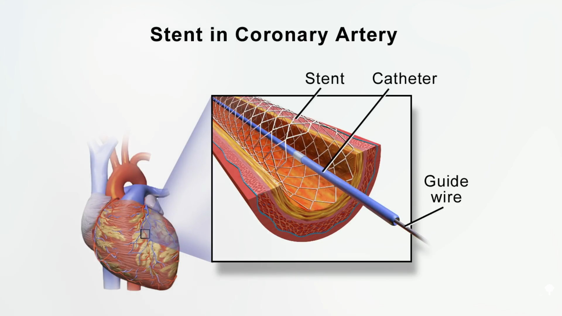
Please participate in the Mass-II trial. This didn’t make any profits even after a year, but no profits even after five or ten years. Then came the trial of courage, which randomized thousands of patients, but it also flattened to the face.
However, although bare metal stents were primarily used, they are not new “drug eluting” stents that release the drug slowly. And what about high-risk groups, such as those diagnosed with diabetes or other more serious illnesses, or those blocking 100% of their arteries after having a heart attack? In the meta-analysis, after seeing five trials in 5,000 patients, there was no reduction in death, heart attack, or even angina pain. Ten trials in over 6,000 patients did not benefit from survival, heart attack, or pain relief. Well, we have done over 12 major exams and there is nothing. There is no benefit from angioplasty or stents. “In addition, multiple analyses failed to identify a single high-risk subset that would benefit…” How is that possible? You are physically open to the bloodstream.
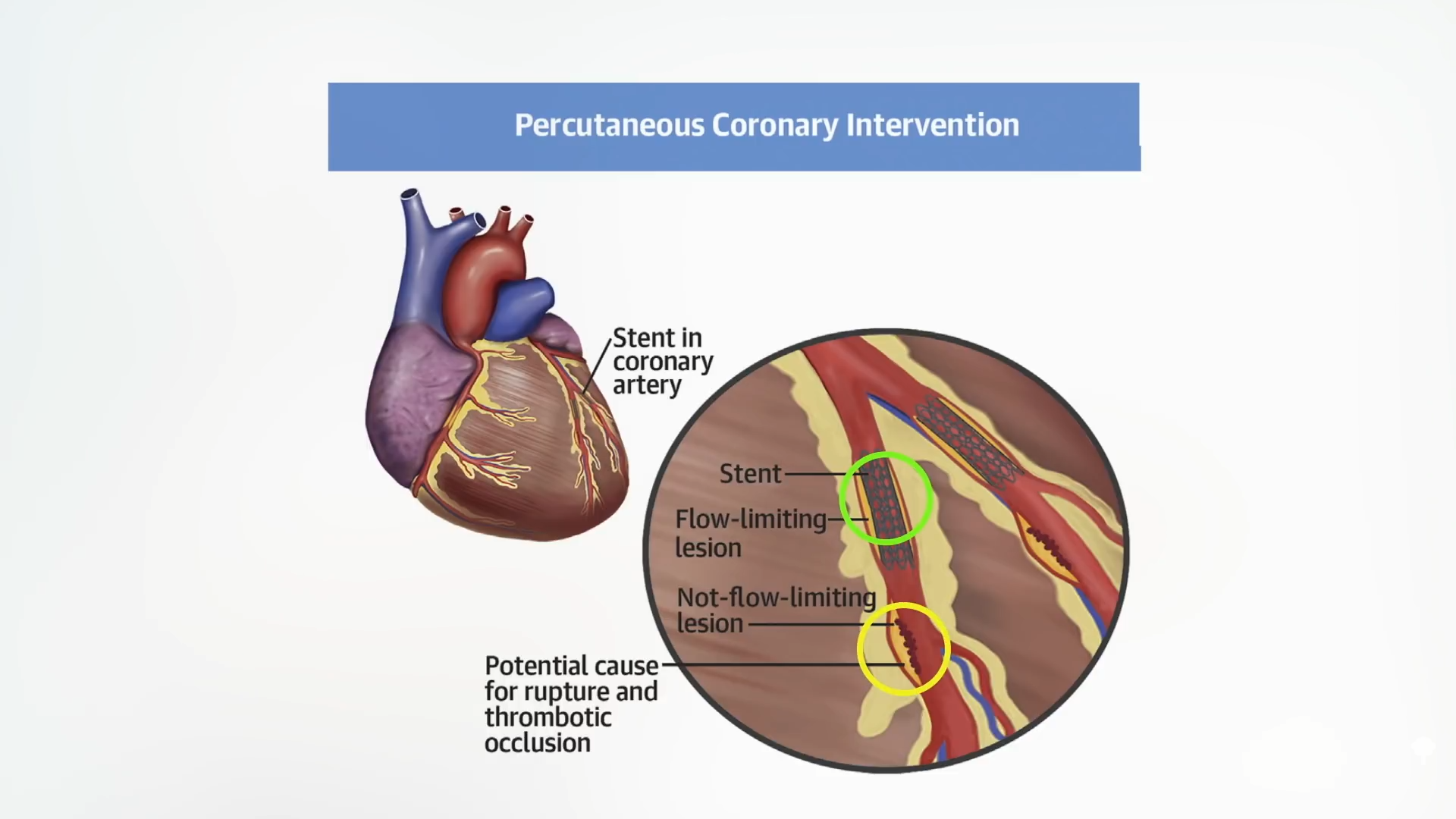
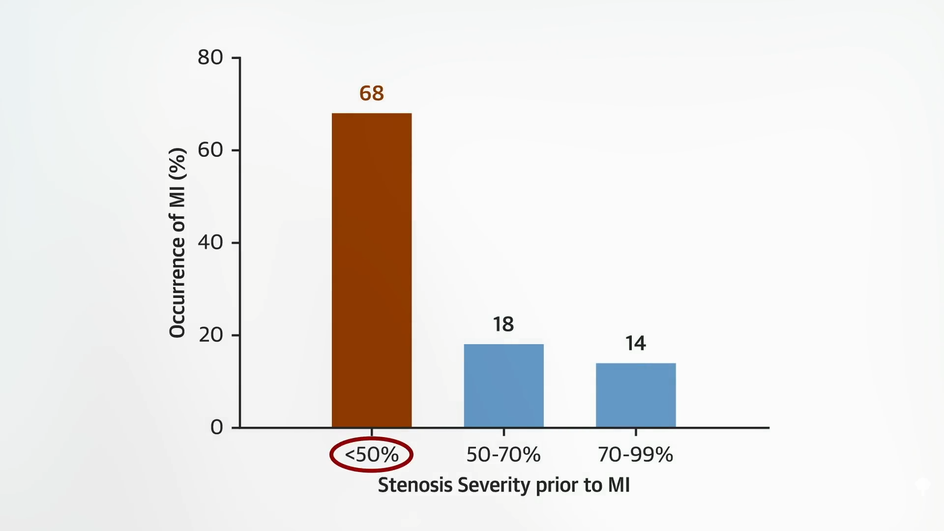
The reason it doesn’t work is because the majority of the heart attacks in real life are less than 70% (i.e., lesions that presumably limit flow) – the stenosis of the arteries that kill us tend to have a tendency to not limit blood flow. My video shows below, and there are two atherosclerotic plaques at 3:21. The ones surrounded by green and labeled “flow-restricting lesion” can very narrow the blood flow and be seen with angiography, and the doctors can drive it out with a stent.
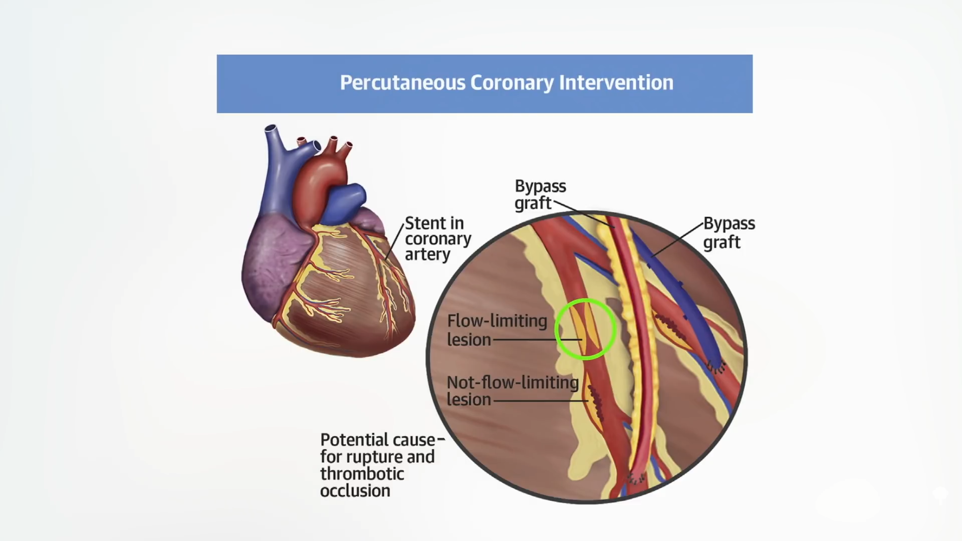
The problem was resolved and life was saved, right? No, as you can see here and at 3:27, it was something invisible (surrounded by yellow below) that didn’t even interfere with the blood flow that had been trying to kill us all the time.
In fact, most heart attacks are caused by non-obstructive plaques that do not reduce blood flow by 50% at 3:40 in my video, as shown below.
There is a misunderstanding. “The analogy of a clogged pipe of stable coronary heart disease (it was particularly difficult,” and cholesterol slowly and relentlessly intrudes blood flow, eventually cutting it off completely, causing a heart attack. In reality, “coronary artery disease is an inflammatory disease in which cholesterol from the blood accumulates on the arterial wall and causes an inflammatory response like acne. When these acne pops, when you inhale blood at the site, these plaques may often be vertically perpendicular and not appear vertically perpendicular, before they burst. Coronary intervention (PCI), i.e., angioplasty and stents. Old plaques are like “dark, old acne.”
The tightest occlusions are mostly composed of dense, fibrous scar tissue, calcified. They can still burst and kill us, but there are many more small lesion brewing hidden from sight. Angiography is the method of visualizing coronary arteries. X-rays are taken after the black-looking dye is injected into the artery, so only the plaque that enters the bloodstream can be seen. That’s why we get these kinds of Iceberg illustrations. The point is to emphasize that most coronary atherosclerotic plaques are not commonly seen by angiography,” as shown in my video at 4:49.

To truly understand what is happening in a person’s artery, we need to look at the autopsy. William Clifford Roberts is perhaps the most outstanding cardiovascular pathologist in the world. What did he learn after 50 years of research into coronary arteries? After examining about 2,000 bodies, he learned that atherosclerosis is a systemic disease.
“In patients with fatal coronary artery disease, the amount of plaque is huge. Here we have not only one plaque, but another plaque with a normal lumen (clean artery) between the plaques. The plaques are continuous! The 5 mm segments do not lack plaques throughout the coronary tree.” Dr. Roberts said: “Segregated coronary artery disease is a myth. There are no such things as “1-vascular disease” or “2-brain disease.” Plaques, if in one, are in all epicardial coronary arteries. ”
The four major coronary arteries nourish the right coronary arteries, the left main coronary arteries, the rotary coronary arteries, and the left anterior descending coronary arteries, as seen at 6:00 in my video.
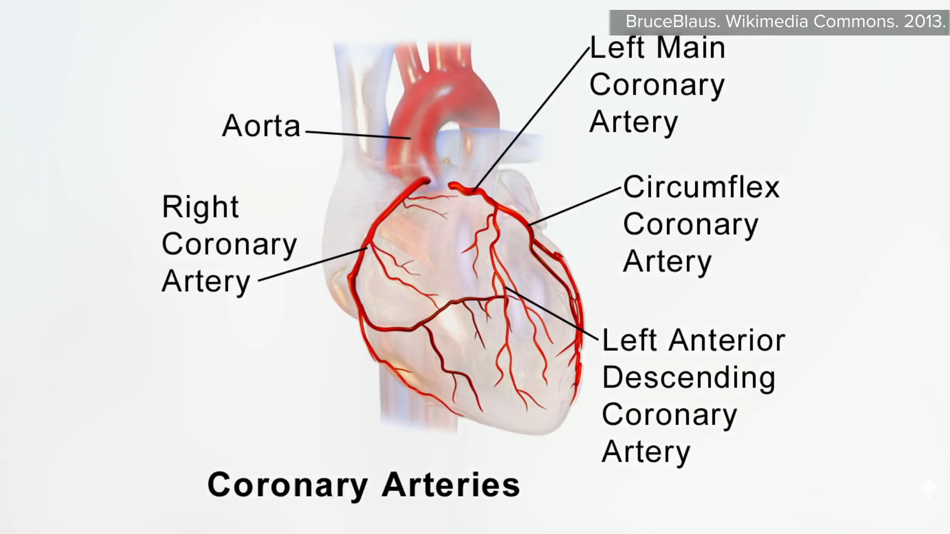
Together, it is a coronary artery of approximately 11 inches (28 cm) and can be cut into approximately 50 inches (5 mm) slices for examination. My video is shown below and can be seen at 6:17. The plaque does not gunking one or two slivers. It can be seen throughout all coronary arteries. Looking at more than 1,000 of these slices from dozens of patients who died from heart attacks, “a single segment lacked plaque.” So it’s no wonder that openness in one area does not affect heart attacks or death.
